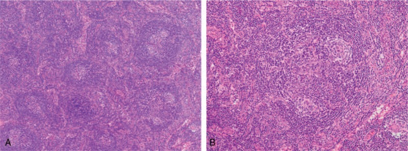
Figure 1.

Histologic findings of the cervical lymph node. (A) Lymph node with proliferated high endothelial venules and small number of plasma cells in the interfollicular zone. Hematoxylin and eosin staining (40× magnification). (B) Atrophic germinal centers with enlarged nuclei of high endothelial cells and expanded mantle zones. Hematoxylin and eosin staining (100× magnification).
