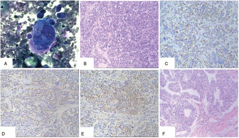
Figure 2.

A, Cytologic examination of ascites showed a large cell. B, The tumor was characterized by small cells with scant cytoplasm, powdery chromatin, and mitotic activity. Tumor cells showed variable arrangements in sheets, clusters, and cords (H&E; original magnification, ×100). C, The ovarian small cell carcinoma was positive for synaptophysin (H&E; original magnification, ×100). D, The ovarian small cell carcinoma was positive for chromogranin A (H&E; original magnification, ×100). E, CKpan positivity was noted in the ovarian small cell carcinoma (H&E; original magnification, ×100). F, The uterus and the left adnexal showed well-differentiated endometrioid adenocarcinoma (H&E; original magnification, ×100).
