
FIGURE 2.
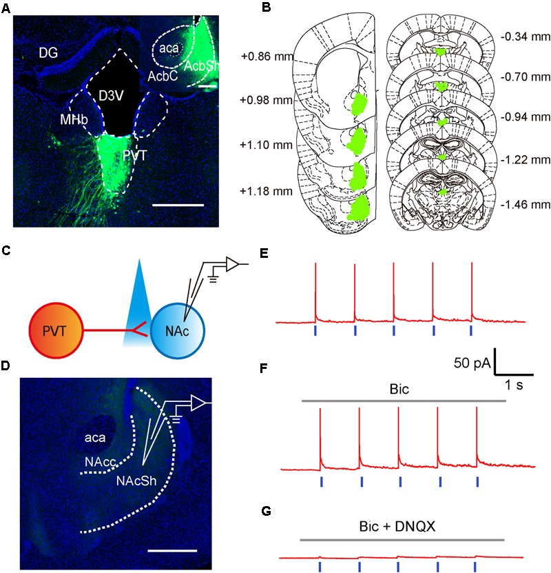
Glutamatergic neurons in the aPVT anatomically and functionally innervate the MSNs in the NAc. (A) Representative confocal image showing the retrograde labeling of retro-AAV2-CMV-GFP (injected in the NAc) in the aPVT. Inset, retro-AAV2-CMV-GFP virus injection site in the NAc. Scale bar = 500 μm. (B) Illustration of the virus infection sites in the NAc and retro-labeled regions in the aPVT overlayed on brain sections adapted from Paxinos and Franklin (2008). Numbers attached to the brain sections indicate the section position posterior to Bregma. (C) Schematic of electrophysiological recording paradigm. (D) Representative confocal image showing the recording site in the slices of mouse brain. Scale bar = 500 μm. (E) Representative trace of action potentials recorded from an MSN evoked by light stimulation at 1 Hz in the NAc. (F,G) Representative traces of action potentials evoked by light stimulation at 1 Hz in the NAc in the presence of bicuculine (bic) (F) or bic and DNQX (G).