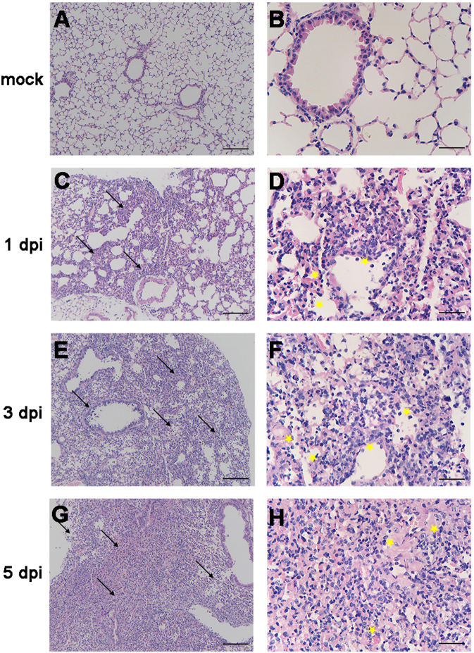
Fig. 2. Histopathology of the lungs from H5N6/GZ14-infected mice.
a, b Eight-week-old female BALB/c mice without infection. c–h Eight-week-old female BALB/c mice infected with 10 MLD50 (50 pfu) of H5N6/GZ14. Lung tissue sections were stained with hematoxylin-eosin and analyzed under a light microscope. At 1 dpi, inflammatory cells (arrows) could be observed around the bronchi (c, d). The airways showed small volumes of exudates with edema fluid mixed with erythrocytes and inflammatory cells (asterisks). e, f At 3f dpi, the lungs had high numbers of inflammatory cells (arrows), bronchial epithelial intracellular edema, and necrosis with necrotic epithelium sloughing into the airway spaces (asterisks). g, h At 5 dpi, severe pulmonary parenchyma consolidation was observed. Increased accumulation of inflammatory cells and necrotic tissue debris (arrows) was observed in the lung parenchyma. The alveoli were completely filled with edema and hemorrhages (asterisks). Scale bars = 100 μm (a, c, e, g) and 25 μm (b, d, f, h)

