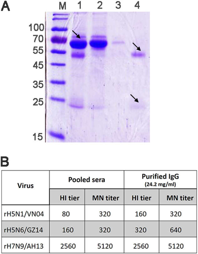
Fig. 4. Coomassie Blue staining after SDS-PAGE and the antibody titer of immunized rhesus macaque sera and purified IgG.

a Coomassie Blue staining after SDS-PAGE of macaque serum and purified IgG. M molecular weight markers (kDa). Lane 1: pooled hyperimmune rhesus macaque serum (diluted 500-fold). The filtered start serum mainly contains albumin (arrow). Lane 2: the flow through pool and unbound material (diluted 50-fold). Albumin and other proteins were removed and could be observed in the flow through pool. Lane 3: the effluent and column wash. Lane 4: the eluted and purified IgG (diluted 1000-fold). The IgG heavy chain (~50 kDa) and light chain (~25 kDa) (arrows) could be observed in the pooled elution buffer without albumin and other proteins. b HI and the MN neutralizing antibody titer of the pooled hyperimmune sera and the purified polyclonal IgG antibody. Three recombinant viruses, rH5N1/VN04, rH5N6/GZ14, and rH7N9/AH13, were used to immunize the rhesus macaques. HI titers are presented as the reciprocal value of the highest serum dilution that inhibited hemagglutination. MN titers are presented as the reciprocal value of the highest serum dilution that conferred 50% neutralization of 100 TCID50 of the virus
