2.
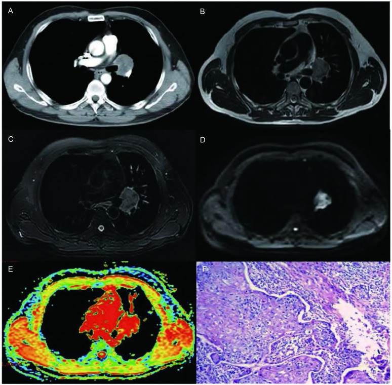
男,51岁,中分化鳞癌。A-E:增强CT、T2WI、T2WI抑脂像、DWI图及ADC图(b=500 s/mm2)。增强CT图显示左肺门软组织肿块,于T2WI及T2WI抑脂像上呈高信号,于DWI图上呈不均匀高信号,于ADC图上以红色区域为主,ADC值为1.67;F:HE染色病理切片(×100)。
A 51-year-old man with moderately differentiated squamous cell carcinoma. A-E:Contrast enhanced CT, T2WI, fat suppressed T2WI, DWI and ADC map (b=500 s/mm2). The contrast enhanced CT shows a tumor in hilum of left lung, and it displays hyperintense in T2WI and fat suppressed T2WI. On DWI, the tumor was inhomogenous hyperintense. On ADC map, the tumor was depicted as an area of red with the ADC value of 1.67; F: histologic sections (100×magnification, H & E staining).

