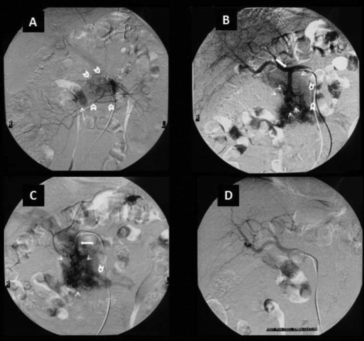
Fig. 5.
(A) Superior mesenteric angiogram depicts the nidus (open arrows), which consists of multiple shunting arterioles. Note early opacification of the portal vein (arrow) in the arterial phase. (B) Celiac angiogram demonstrating dilatation of the gastroduodenal artery (arrow) and a tightly packed mass of enlarged feeding arteries, which represent the pancreatic arteriovenous malformation (small arrows). Note the visible draining vein (open arrows). (C) Selective angiogram of the gastroduodenal artery shows that it is the main feeding artery. Note the early opacification of the superior mesenteric vein and of the portal vein. (D) Celiac angiogram post PVA embolization of the main feeding arteriole and after additional coil placement, showing a reduction of the nidus size and no early venous return to the portal system.

