
Figure 5.
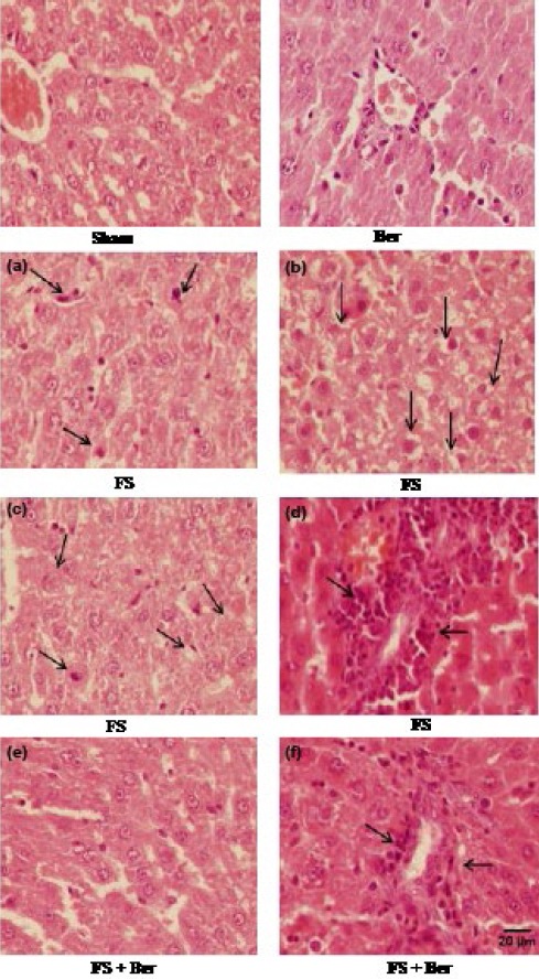
Representative light microphotographs of the livers. Haematoxylin and eosin-stained sections were evaluated by light microscopy. The histological changes observed included apoptosis (a), swelling (b), vacuolization (c), and inflammatory cell infiltration (d)