
Figure 6.
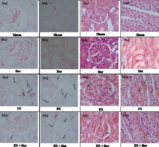
Representative light microphotographs of the kidneys. Prussian blue- and hematoxylin & eosin-stained sections were evaluated by light microscopy. The histological changes observed included iron deposition in the cytoplasm of: cortical (c1, d1) and medullary (c2, d2) tubular cells, Prussian blue staining; glomerular capillary congestion (c3, d3) and medullary vascular congestion (c4, d4), haematoxylin-eosin staining