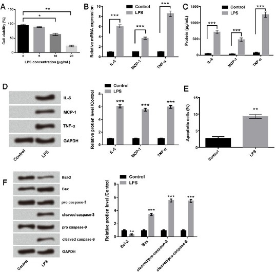
Figure 1.

Lipopolysaccharide (LPS) induced inflammatory damage in WI-38 cells. (A) The effect of LPS with different concentrations (5, 10, and 20 μg/ml) on cell viability was analyzed by CCK-8 assay. The expression levels of IL-6, MCP-1, and TNF-α were analyzed by (B) qRT-PCR, (C) ELISA, and (D) Western blot after treatment with 10 μg/ml LPS. The effect of LPS with 10 μg/ml on cell apoptosis was evaluated by (E) Annexin V-FITC/PI double staining method and (F) Western blot. Means ± SD were shown. * P<0.05, ** P<0.01 *** P<0.001 (ANOVA)
