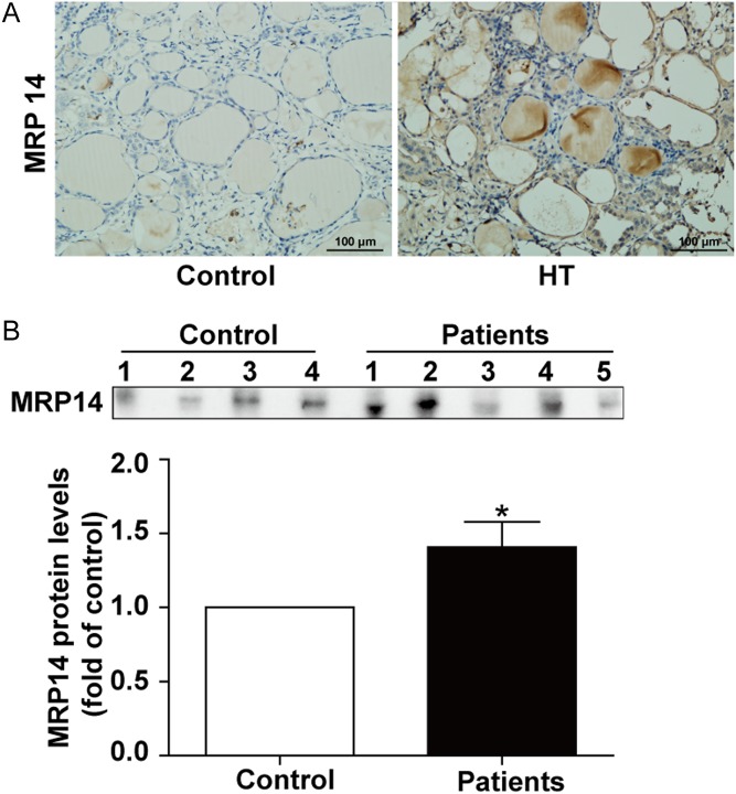
Figure 1.
MRP14 expression in the thyroid tissues and sera of Hashimoto’s thyroiditis (HT) patients. (A) Representative results of MRP14 immunohistochemical staining in HT tissues (n = 6) and control tissues (n = 5) are shown. ‘Control’ indicates tissues from patients with simple goitre of the thyroid. Brown regions represent positive expression (original magnification, ×200; scale bars, 100 μm). (B) Serum MRP14 levels of HT (n = 20) and healthy controls (n = 20) were analysed by immunoblots. Representative immunoblotting results of MRP14 (upper panel) are shown. The results of immunoblot quantification from all samples are shown (lower panel). Significant differences and P values are calculated by unpaired t-tests. *P < 0.05 vs controls.

 This work is licensed under a