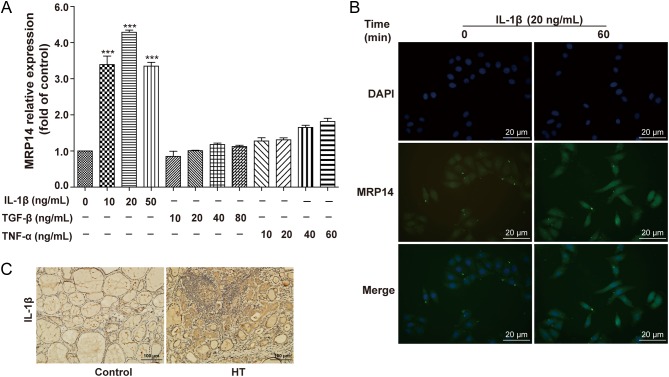
Figure 2.
IL-1β induces MRP14 expression in Nthy-ori 3-1 cells. (A) Nthy-ori 3-1 cells were starved overnight and then incubated with gradient concentrations of IL-1β, TGF-β and TNF-α for 4 h, and cells were harvested for quantitative real-time PCR (qPCR). The results shown are representative of three replicates (left upper panel). (B) Nthy-ori 3-1 cells were starved overnight and then incubated with IL-1β 20 ng/mL. MRP14 levels were detected by immunofluorescence staining (original magnification, ×400; scale bars, 20 μm). (C) Representative results of IL-1β immunohistochemical staining in HT tissues (n = 4) and control tissues (n = 4) are shown (original magnification, ×200; scale bars, 100 μm). Significant differences and P values were calculated by one-way ANOVA and the Mann–Whitney U tests. ***P < 0.001 vs controls.

 This work is licensed under a