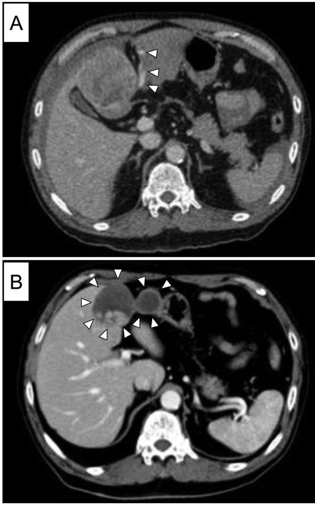
Figure 1. A: Portal phase of enhanced computed tomography at the time of spontaneous tumour rupture. A 6-cm hepatocellular carcinoma was present in segment 4 of the liver, and fluid collection was present in the peritoneal cavity. The arrowheads indicate extravasation of haemorrhage. B: Enhanced computed tomography 3 months after transcatheter arterial embolization revealed a reduction in the volume of the main tumour and intra-abdominal haemorrhage (arrowheads) and no intrahepatic metastasis or peritoneal dissemination. Adhesion between the remaining haemorrhage and stomach was suspected.