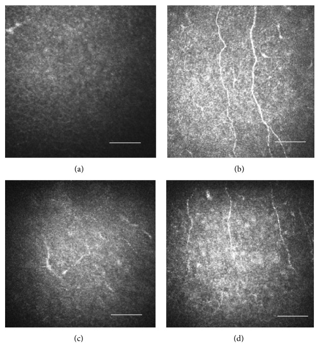
Figure 2.
Evaluation of the subbasal nerve fibers in the cornea with the HRT II RCM. Size bar = 100 μm. (a) Four months after the nerve block, the subbasal nerve fibers were hardly observed in the right eye. (b) Four months after the nerve block, the subbasal nerve fibers were observed in the fellow eye. (c) Six months after the nerve block, regenerated subbasal nerve fibers were observed starting from the periphery of the right cornea, although they appeared small and short. (d) Six months after the nerve block, subbasal nerve fibers were observed in the fellow eye.

