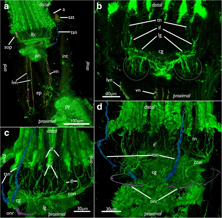
Fig. 11.
Innervation of the lophophoral base in Cinctipora elegans. CLSM, staining of acetylated alpha-tubulin (green) and serotonin-lir nervous system (in A, yellow). a Lateral view of the polypide showing the visceral neurite bundles (highlighted in red) and tentacle sheath neurite bundle (highlighted in light blue). b View of the anal side of the cerebral ganglion and the lateral ganglia which are proximally delineated by a dashed line. The visceral neurite bundles are highlighted in red. c Oblique view of the lophophoral base showing the circum-oral nerve ring, the tentacle nerves, the tentacle sheath nerves (highlighted in light blue) and the (incomplete) outer ring nerve (highlighted in purple). d View of the anal side showing the ganglion with the tentacle sheath nerves (highlighted in light blue) and the (incomplete) outer ring nerve (highlighted in purple). Abbreviations: a – anus, cg – cerebal ganglion, ep – esophagus, if – intertentacular fork, int – intestine, lb – lophophoral base, lf- latero-frontal nerve of tentacle, lg – lateral ganglion, lvn – laterovisceral neurite bundle, mf – medio-frontal nerve of tentacle, onr – outer nerve ring, ph – pharynx, py – pylorus, sat – serotonin-lir anal tube, sop – serotonin-lir oral perikarya, tn – tentacle nerves, tsn – tentacle sheath nerve, vn – visceral nerves

