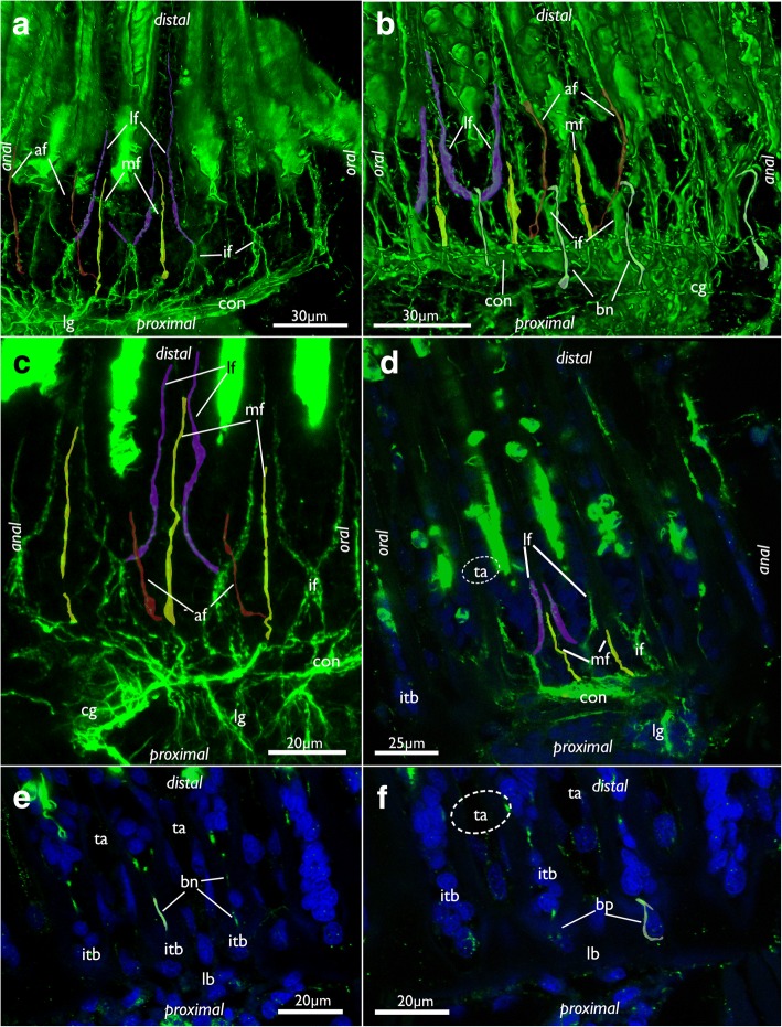
Fig. 14.
Tentacle innervation in Cinctipora elegans. CLSM, staining of acetylated alpha-tubulin (green) and cell nuclei (DAPI, blue). Some mediofrontal neurite bundles are highlighted in yellow, latero-frontal neurite bundles in purple and abfrontal neurite bundles in red. The basal neurite bundle is highlighted in white. a Lateral view of the main tentacle neurite bundles projecting from the circum-oral nerve ring. Volume rendering. b Similar view as in A but showing more lateral portions of the lophophore including the basal neurite bundle which originates from the intertentacular fork and extends proximally into the intertentacular base. c Projection images of few optical sections showing the tentacle innervation in detail. d Optical section of the intertentacular fork and the medio- and latero-frontal nerves. e Optical section of the lateral side of the lophophoral base showing basal radial neurite bundles. f Optical section of the same specimen as in E, but more superficially showing the basal perikarya at the proximal end of the intertentacular bases. Abbreviations: af – abfrontal neurite bundle of the tentacle, bn – basal neurite bundle, bp – basal perikarya, cg – cerebral ganglion, con – circum-oral nerve ring, if – intertentacular fork, itb – intertentacular base, lb – lophophoral base, lf - latero-frontal neurite bundle of the tentacle, lg – lateral ganglia, mf – mediofrontal neurite bundle of the tentacle, ta – area where tentacles extend distally

