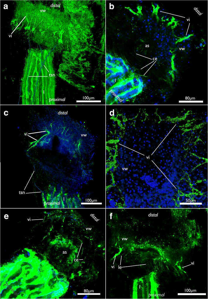
Fig. 16.
Nervous system of the apertural area of Cinctipora elegans, CLSM, stainings of acetylated alpha-tubulin (green) and cell nuclei (DAPI, blue). a Lateral view of a volume rendering of the apertural area. Tentacle sheath nerves in the proximal part are highlighted in light blue. Volume rendering. b Close-up showing circular neurite bundles in the extracellular matrix of the attachment organ. Volume rendering. c Optical section through the vestibular wall. Tentacle sheath neurite bundles in the proximal part are highlighted in light blue. d View from the distal side onto the apertural area showing radial arrangement of the vestibular innervation. Volume rendering. e Oblique view with circular neurite bundles in the extracellular matrix of the attachment organ. Volume rendering. f Optical section showing the thicker neurite bundles in the vestibular wall compared to the longitudinal bundles within the extracellular matrix. Abbreviations: as – atrial sphincter, ce - circular neurite bundles in the extracellular matrix of the attachment organ, l – lophophore, le - longitudinal neurite bundles in the extracellular matrix of the attachment organ, tsn – tentacle sheath nerves, vi – vestibular innervation, vw – vestibular wall

