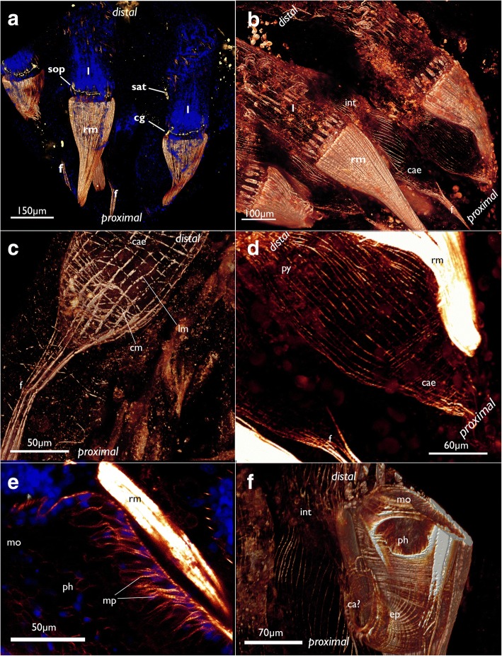
Fig. 19.
Myoanatomy of the digestive tract of Cinctipora elegans. CLSM, staining of f-actin (phalloidin, glow-LUT) and cell nuclei (DAPI, blue). a Overview of several polypides showing the prominent retractor muscles that cover most parts of the foregut. The serotonin-lir nervous system is displayed in yellow. Volume rendering. b Similar as in A with higher magnification of some zooids showing again the prominent retractor muscle covering the foregut and the loosely arranged, mostly thin, longitudinal musculature of the stomach (caecum, pylorus) and hindgut (intestine). Volume rendering. c Detail of the proximal end of the caecum showing few circular muscle fibers next to the more prominent longitudinal ones. The latter extend proximally into the longitudinal musculature of the funiculus. Volume rendering. d Overview of the stomach with the proximal caecum and the more distally located pylorus. e Optical section of the foregut showing the myoepithelial cells of the pharynx. f Detail of the foregut showing its dense circular as well as longitudinal muscle bundles in the esophagus towards the stomach. Volume rendering. Abbreviations: ca? – area corresponding the cardiac area of the stomach in other bryozoans, cae – caecum, cg – cerebral ganglion, cm – circular muscles of the caecum, ep – esophagus, int – intestine, l – lophophore, lm – longitudinal muscles of the caecum, mo – mouth opening, mp – myoepithelial cells of the pharynx, ph – pharynx, py – pylorus, rm – retractor muscle, sat – serotonin-lir anal tube, sop – serotonin-lir oral perikarya

