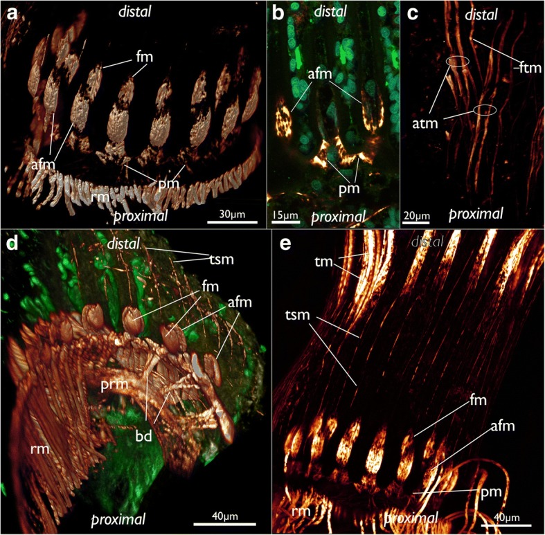
Fig. 21.

Musculature of the lophophoral base and the lophophore in Cinctipora elegans. CLSM, staining of f-actin (phalloidin, glow-LUT), acetylated alpha-tubulin (green) and cell nuclei (DAPI, cyan). a Oblique view of the lophophoral base showing the lophophoral base muscles. Volume rendering. b Optical section through the lophophoral base musculature. c Projection image of several optical sections of the tentacle muscles. Note that two separate bundles may occur on the abfrontal side. d Oblique view of the mouth opening and the lophophoral base showing the short buccal dilatators. Volume rendering. e Lateral view of the lophophoral base including tentacle sheath muscles. Note also the gap from the lophophoral base muscles until the tentacle muscles appear. Abbreviations: afm – abfrontal muscles of the lophophoral base, atm - abfrontal tentacle muscle, bd – buccal dilatators, fm – frontal muscles of the lophophoral base, ftm - frontal tentacle muscle, pm – proximal lophophoral base muscles, prm – pharyngeal ring muscles, rm – retractor muscle, tm – tentacle muscles, tsm – tentacle sheath muscles
