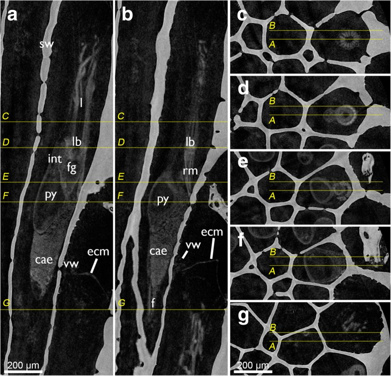
Fig. 3.

Tomographic sections of skeleton and soft-body visualization by microCT of Cinctipora elegans. a and b are longitudinal sections of two zooids showing the homogenous skeletal wall and the lighter shades of grey of the polypide. The yellow lines indicate cross-sectional planes displayed in c-g which progress from distal to proximal. Abbreviations: cae – caecum, ecm – thick extracellular matrix of the attachment organ, f – funiculus, fg – foregut, int – intestine, l – lophophore, lb. – lophophoral base, py – pylorus, rm. – retractor muscle, sw – skeletal wall, vm – vestibular wall
