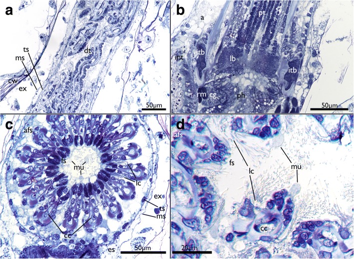
Fig. 7.
Histological details of the tentacles of Cinctipora elegans. Semithin sections, toluidine blue. a Longitudinal section of the retracted tentacle crown. The distal part of the tentacles are contorted and twisted due to contraction of the tentacle muscles. b Longitudinal section through the proximal part of the lophophoral base and proximal parts of the tentacles. The latter are not contorted in this area. c Cross-section through the proximal area of the lophophore showing the regular circular arrangement of the tentacles. Note also globular secretions, probably mucus, on the frontal side of the tentacles and enclosed within the lophophore. d Cross section through distal parts of the tentacles showing its more contorted form and secretory droplets being formed on their frontal side. Abbreviations: a – anus, afs – abfrontal side of tentacles, cc – central cells, cg – cerebral ganglion, cw – cystid skeletal wall, dt – distal part of tentacles, es – endosaccal space (coelom), ex – exosaccal space, fs – frontal side of tentacles, int – intestine, itb – intertentacular base, lb – lophophoral base, lc – lateral cilia, ms – membranous sac (peritoneum), mu – mucus-like secretions, ph – pharynx, pt – proximal part of tentacles, rm – retractor muscle, ts – tentacle sheath

