Figure 4.
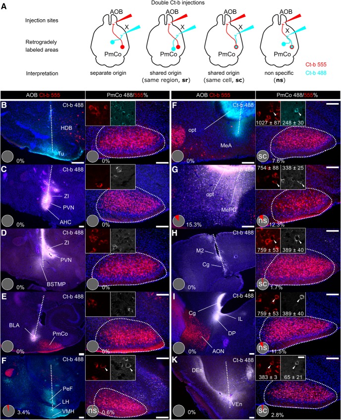
Collateral projections of PmCo corticobulbar neurons. A, Dual Ct-b injections were used to identify possible additional target areas of the PmCo neurons retrogradely labeled from the AOB (Ct-b 555). Tracing was considered reliable in case of clear separation of the two injection sites and 555/488 colabeling of the same region (sr) or the same cells (sc). F–I, We considered nonspecific (ns) tracing experiments to be those in which the two tracers showed partial overlap near the two injection sites or in case of Ct-b 488 injections adjacent to the stria terminalis (for reference, see Allen Brain Connectivity Atlas, experiment #114249084) where AOB-directed ACPs course (e.g., G, I): in such cases, Ct-b 488 would be likely taken up by passing fibers and yield false-positive results (compare H to I and G to F to see how Ct-b overlap in the PmCo decreases as the injection site is moved either dorsally or rostrally, respectively). B–H, Injections of green Ct-b 488 (cyan) were targeted to different AOS regions, known main targets of the PmCo: the olfactory tubercle (Tu; B), the paraventricular nucleus (PVN; C, D), the bed nucleus of the stria terminalis (BNST; D), the ventromedial hypothalamic nucleus (VMH; E), the MeA (G; MePD, H), the basolateral amygdaloid nucleus (BLA, F), the endopiriform nuclei (K), and the medial prefrontal cortex were targeted (I, J). The pie charts in the panels showing the injections sites (left) indicate the coexpression of Ct-b 555 in Ct-b 488 fibers and therefore possible biases due to nonspecific tracing (tracing reliability is indicated according to the diagrams in A). Similarly, the coexpression of Ct-b 488 and Ct-b 555 in the PmCo is indicated in percentage in the panels on the right. In the regions showing higher coexpression, the absolute (averaged) values are indicated in the high-magnification insets. HDB, Nucleus of the diagonal band of Broca; ZI, zona incerta; BSTMP, bed nucleus stria terminalis medial division posterior part; PeF, perifornical nucleus; LH, lateral hypothalamic nucleus; opt, optic tract; M2, secondary motor cortex; Cg, cingulate cortex; IL, infralimbic cortex; DP, dorsal peduncular cortex; Den, dorsal endopiriform nucleus; VEn, ventral endopiriform nucleus. Scale bars: left panels, 20 μm; right panels, 200 μm. Data are the mean ± SEM.

