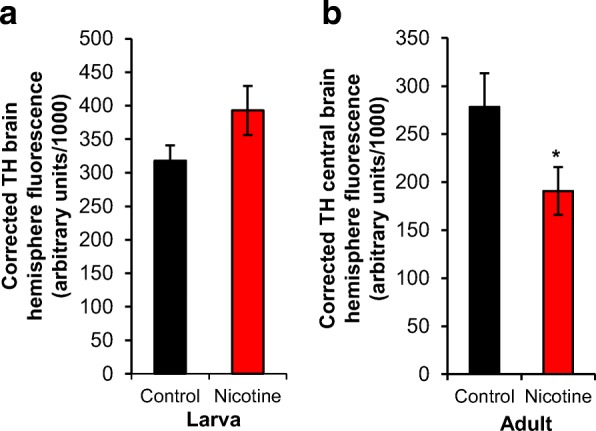
Fig. 3.

Developmental nicotine exposure decreases TH fluorescence in adult brains. Flies were raised on control food or food laced with 0.3 mg/ml nicotine. Larvae were dissected at the 3rd instar stage of development; adults were dissected 4 days after eclosion. The brains were stained with an antibody against tyrosine hydroxylase (TH). a Corrected TH brain hemisphere fluorescence, which normalized the staining fluorescence to background levels, showed that developmental nicotine exposure had no statistically significant effect on TH staining in larval brains. b Corrected TH central brain fluorescence was significantly decreased in adult brains of flies exposed to nicotine during development. Sample size was n = 22 brain hemispheres for control and n = 18 brain hemispheres for nicotine from 5 independent experiments for the larval stage, and n = 10 brains for control and n = 17 for nicotine from 5 independent experiments for adult. Mann-Whitney U-test (a) and Student’s t-test (b) were used to compare the control versus the nicotine condition
