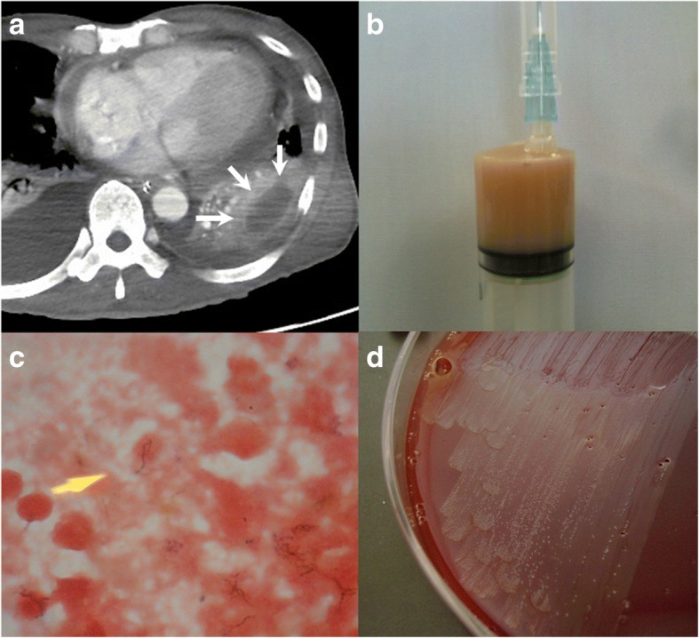
Fig. 3.

a The chest CT with enhancement on the 9th day showed a thick-walled cavitary lesion containing water density in the left lower lobe (white arrows) and a small amount of pericardial effusion; b Purulent fluid obtained by ultrasound-guided pneumocentesis of the cavitary lesion in the left lower lobe; c Gram stain of the fluid showed Gram-positive filamentous rods; d Cultures from the fluid grew Actinomyces species
