. 2018 May 16;9(23):5212–5222. doi: 10.1039/c8sc01262h

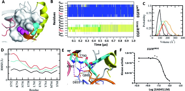
Fig. 4. Characterization of the druggable allosteric pocket in EGFRL861Q: (A) structure of the EGFRWT kinase in its inactive form with the αC-helix (yellow), short α-helix (orange) and the hydrophobic allosteric pocket shown in surface with buried residues Leu858 (shown as magenta spheres) and Leu861 (shown as cyan spheres); (B) secondary structure evolution (blue: α-helix, yellow: β-strand, gray: 310-helix, green: turn, and white: coil) of the short α helix (y-axis) during the MD simulations of EGFRWT and EGFRL861Q. (C) Volume of the allosteric pocket calculated from the conformations sampled during the MD simulations of EGFRWT (black), EGFRL858R (red), EGFRL861Q (cyan), and EGFRL858R/T790M (orange). (D) RMSF of the αC-helix during the MD simulations of EGFRWT (black), EGFRL858R (red) and EGFRL861Q (cyan). (E) Snapshot from the MD simulation of the complex between EGFRL861Q and EAI045 (cyan carbon, stick representation). All the interacting residues are highlighted as thin sticks and all the interactions are highlighted as dotted lines (magenta). Colouring of the kinase structure is the same as in Fig. 1. (F) Binding affinity (Kd) of EAI045 for EGFRL861Q measured experimentally using KINOMEscan™ at DiscoverX.

Fig. 4