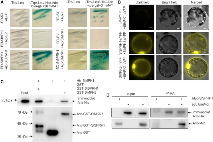
Figure 5.
Interactions between SlMPK1 and SlSPRH1. A, Y2H assay of interactions between SlMPK1 and SlSPRH1. SlMPK1 (as bait) was cloned into pGBKT7 (BD) and SlSPRH1 (as prey) was cloned into pGADT7 (AD). AD-T and BD-p53 was used as a positive control, and AD-empty vector (EV) and SlMPK1-BD was used as a negative control. Simultaneously, SlSPRH1 (as bait) was cloned into pGBKT7 (BD) and SlMPK1 (as prey) was cloned into the pGADT7 (AD). AD-SlMPK1 and BD-EV was used as a negative control. B, BiFC assay for detecting molecular interactions between SlMPK1 and proteins (SlMKK2 or SlSPRH1) transiently coexpressed in tobacco leaf protoplasts. SlMPK1 was fused with the C terminus of YFP, and SlMKK2 and SlSPRH1 were fused with the N terminus of YFP. The images were obtained from the YFP channel, the differential interference contrast channel, and a merged image of the two channels. The positive control was SlMKK2-nYFP/SlMPK1-cYFP, and the negative control was EV-nYFP and SlMPK1-cYFP. Bars = 50 μm. C, SlMPK1 interacts physically with SlSPRH1. His-tagged SlMPK1 was incubated with immobilized GST or GST-tagged SlSPRH1. Beads were washed, fractionated by 12% (w/v) SDS-PAGE, and subjected to immunoblot analysis using an antibody against His (top) or GST (bottom). Immobilized GST was used as a negative control, and GST-tagged SlMKK2DD was used as a positive control. D, In vivo Co-IP assay of the SlMPK1 and SlSPRH1 interaction in tobacco leaves. Crude lysates precleared by protein A-Sepharose beads (In-put) were immunoprecipitated (IP) with anti-HA antibody and then detected with anti-HA and anti-Myc antibodies for SlMPK1-HA and SlSPRH1-Myc, respectively.

