Figure 7.
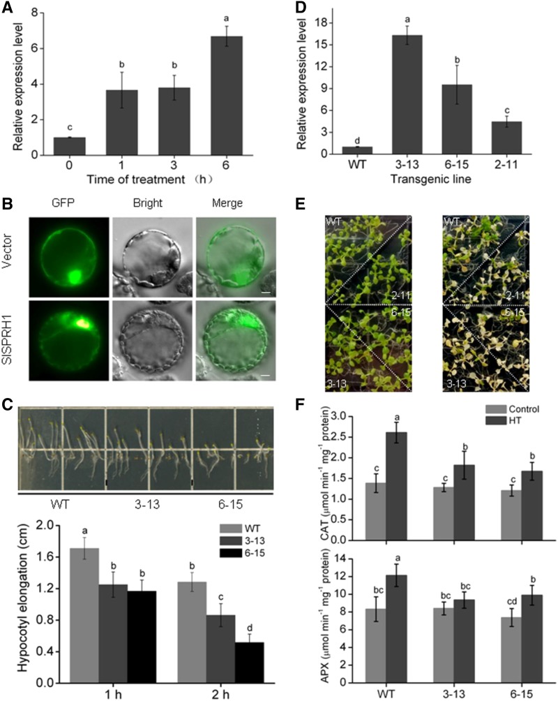
SlSPRH1 negatively regulates Arabidopsis tolerance to HT stress. A, SlSPRH1 expression is induced by HT. RT-qPCR analyses of transcript abundance of SlSPRH1 is shown in leaves of wild-type tomato plants during HT (42°C) for 0, 1, 3, and 6 h. B, Subcellular localization of SlSPRH1. The SlSPRH1-GFP protein and GFP, driven by CaMV 35S, were transformed separately into tobacco protoplast and visualized by fluorescence microscopy. Images were taken in representative cells expressing GFP (top) or SlSPRH1-GFP fusion protein (bottom) under bright field (middle) or dark field (left). The merged images are shown at right. Bars = 50 μm. C, Hypocotyl length of wild-type (WT) and SlSPRH1-expressing Arabidopsis lines in one-half-strength Murashige and Skoog medium under HT stress. After germination for 3 d, the plates covered with foil were subjected to HT treatment at 45°C for 1 and 2 h, followed by vertical culturing in the dark for 6 d. D, Expression of SlSPRH1 in SlSPRH1-expressing lines. RT-qPCR analyses of the transcript abundance of SlSPRH1 is shown in the three independent transgenic plants. ACTIN was used as an internal control. E, Phenotypes of SlSPRH1-expressing transgenic lines 3-13, 6-15, and 2-11 under HT stress. Ten-day-old Arabidopsis seedlings in one-half-strength Murashige and Skoog medium (left) were subjected to HT treatment at 42°C for 1.5 h, followed by recovery for 10 d (right). F, Response of antioxidant enzymes to HT in the SlSPRH1-expressing plants. Two-week-old wild-type and transgenic line 3-13 and 6-15 plants were treated with HT for 3 h. The activities of CAT and APX were determined. Error bars represent sd values of three replicates. Statistical differences among the samples are labeled with different letters according to the lsd test (P < 0.05, one-way ANOVA).

