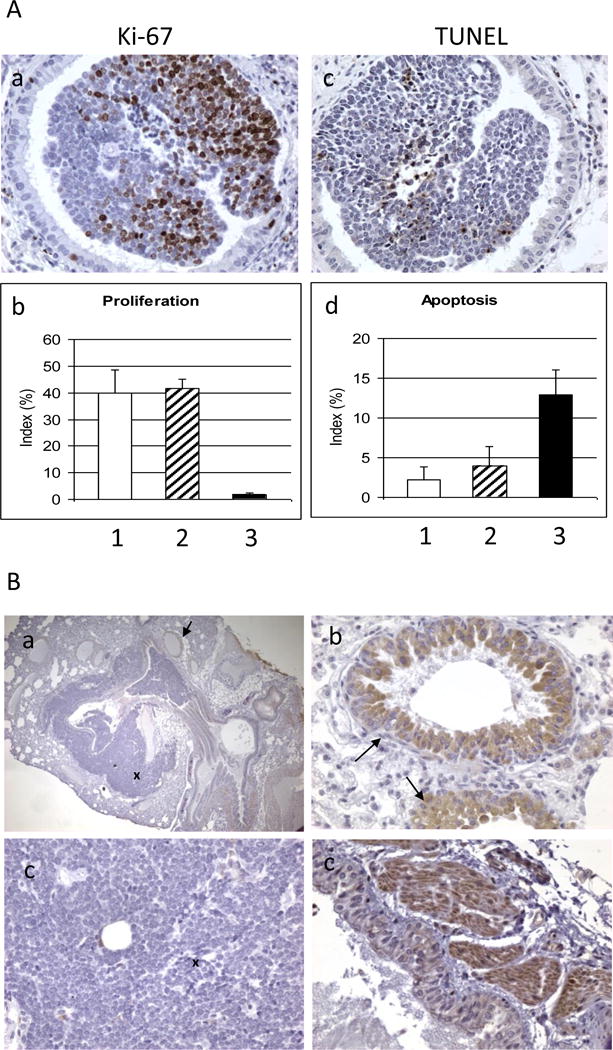
Figure 3. IHC staining.

A. Effect of bexarotene on cell proliferation and apoptosis. Representative photo images of Ki-67 stains (a) and TUNEL stains (c) are shown. Bexarotene decreased cell proliferation (b; 1, control; 2, budesonide; 3, bexarotene) and increased apoptosis in mSCLC (d; 1, control; 2, budesonide; 3, bexarotene). B. IHC staining on mSCLC in B5 (AJ × Trp53F2-10/F2-10;Rb1F19/F19) mice with anti-glucocorticoid receptor (GR) antibody. Panel a shows positive staining of GR in bronchial epithelial cells (indicated by arrow) and negative staining in SCLC cells (indicated by cross); Panel b shows enlarged bronchial epithelial cells; Panel c shows enlarged SCLC cells; and Panel d shows the positive staining in muscle cells). Original magnification 100× in panel a; 400× in panels b-d.
