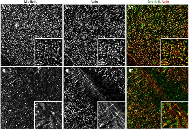
Fig. 9.
Mid1ip1L protein localizes with cortex and furrow actin. (A-B″) SIM images of single and merged channels showing partial colocalization of Mid1ip1L protein with F-actin in wild-type embryos. (A-A″) At the cortex, Mid1ip1L protein partially colocalizes with the cortical F-actin. (B-B″) At the furrow, Mid1ip1L protein also partially colocalizes with transverse F-actin at furrow distal ends, which corresponds to furrow F-actin indentations (see also Fig. S5). Insets show a magnified view within the field. Scale bar: 10 μm in A-B″; 1 μm in insets.

