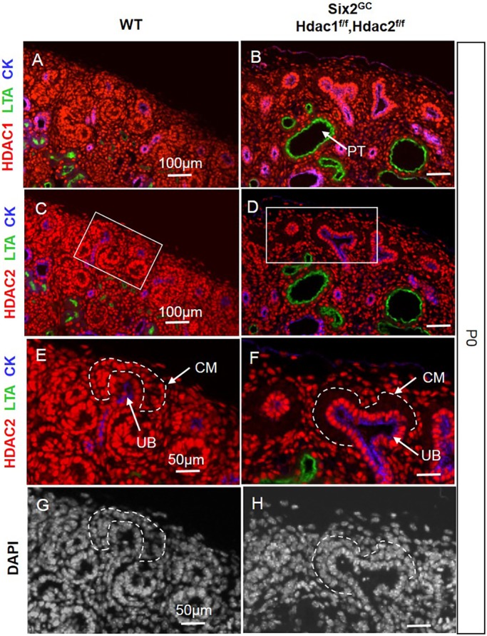
Fig. 1.

Deletion of Hdac1 and Hdac2 genes in the CM. (A,C,E,G) Consecutive section immunofluorescence (IF) at P0 showing the relatively abundant nuclear expression of HDAC1/2 proteins in the nephrogenic zone within the CM, UB and stroma. (B,D,F,H) Conditional Six2-Cre-mediated deletion of Hdac1/2 genes results in efficient loss of HDAC1/2 proteins from the CM. Boxes in C and D are shown enlarged in E and F, respectively. The scale bar information is the same in the left-hand and right-hand panels. CK, pancytokeratin; CM, cap mesenchyme; LTA, Lotus tetragonolobus lectin; PT, proximal tubule; UB, ureteric bud branches.
