Fig. 3.

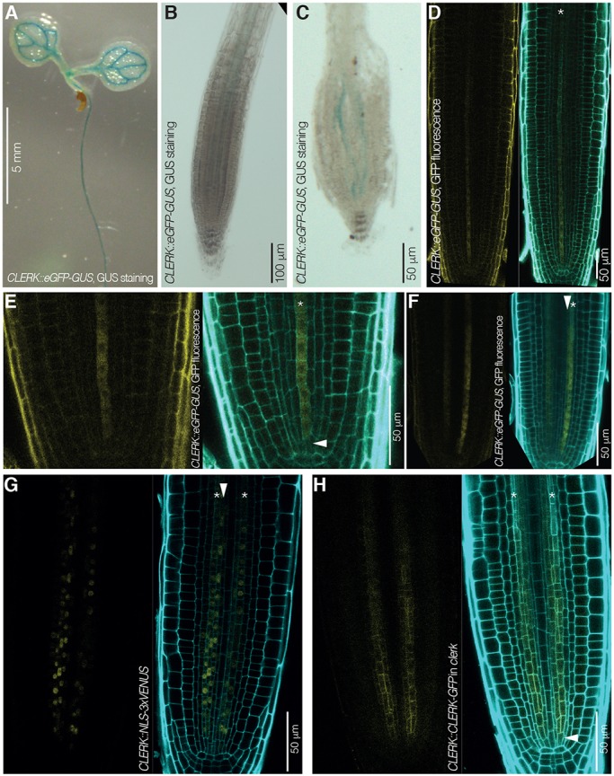
CLERK expression pattern in Arabidopsis seedlings and roots. (A-C) CLERK expression pattern revealed by GUS staining (blue) in 7-day-old Col-0 seedlings that express an eGFP-GUS fusion under control of the CLERK promoter (CLERK::eGFP-GUS); light microscopy. (A) Cotyledons and upper part of the root. (B) Higher magnification of the root tip. (C) Higher magnification of the root tip after squashing, revealing expression in two cell files. (D-F) CLERK gene expression pattern revealed by GFP fluorescence in root meristems of 7-day-old Col-0 seedlings that carry a CLERK::eGFP-GUS transgene; confocal microscopy. Left: GFP fluorescence, yellow. Right: overlay of GFP fluorescence with propidium iodide (PI) cell wall staining (cyan). (D) Root tip overview. Asterisk indicates a protophloem sieve element cell file. (E) Similar to D, higher magnification of the meristem tip, highlighting CLERK expression in early protophloem (asterisk) starting next to the protophloem stem cell (arrowhead). (F) Similar to D, alternative perspective angle, highlighting CLERK expression in early protophloem sieve elements (asterisk) as well as metaphloem cells (arrowhead). (G) CLERK expression visualized by VENUS fluorescence in root meristems of 7-day-old Col-0 seedlings that carry a CLERK::NLS-3×VENUS transgene, confocal microscopy. Left: VENUS fluorescence, yellow; Right: overlay of VENUS fluorescence with PI cell wall staining (cyan). Asterisks indicate protophloem sieve element cell files; arrowhead indicates a metaphloem cell file. (H) CLERK protein expression pattern revealed by GFP fluorescence in root meristems of 7-day-old clerk-3 seedlings that carry a CLERK::CLERK-GFP transgene; confocal microscopy. Left: GFP fluorescence, yellow. Right: overlay of GFP fluorescence with PI cell wall staining (cyan). Asterisks indicate protophloem sieve element cell files; arrowhead indicates a phloem stem cell.
