
Figure 4.
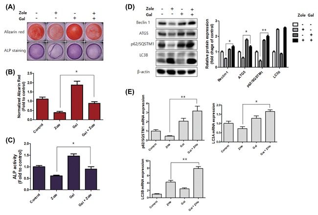
Galangin activates osteoblast differentiation and autophagy‐related factors. A‐C, The hFOB 1.19 cells were pretreated with galangin (5 µM) for 24 h, then treated with zoledronate (50 µM) for 48 h; after treatment, the cells were incubated for 14 days in differentiation‐inducing media. A, Alizarin Red S (upper panel) and ALP staining (lower panel) were conducted to find changes in mineralization and differentiation. The graph indicates the quantification of Alizarin Red S staining (B) and ALP activity (C). D and E, The hFOB 1.19 cells were pretreated with galangin (5 µM) for 24 h, then treated with zoledronate (50 µM) for 48 h. D, Western blotting used antibodies specific for beclin‐1, ATG5, SQSTM1/p62, LC3, and β‐actin. β‐actin was used as an internal control. E, The gene expressions of SQSTM1/p62, LC3A, and LC3B were analyzed by real‐time PCR. The data are expressed as mean ± SD (n = 3) and were analyzed by one‐way ANOVA using Dunnett's multiple‐comparison test. (*P < 0.05 and **P < 0.01 for the difference between the zoledronate and Galangin + zoledronate group)