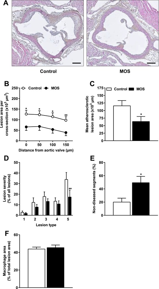
Figure 1.

MOS decreased atherosclerosis development. Mice were fed a WTD with or without MOS for 14 weeks. A) Representative cross sections of the valve area of the aortic root stained with HPS are shown. Scale bar, 200 μm. B) Atherosclerotic lesion area was determined as a function of distance (50 μm intervals) starting from the appearance of open aortic valve leaflets covering 150 μm. C) The mean atherosclerotic lesion area was determined from the four consecutive cross sections, D) lesions were categorized according to lesion severity (type 1–5), E) the percentage of non‐diseased segments were scored, and F) macrophage area within the atherosclerotic lesions were quantified. Open bars/circles represent the control group and closed bars/circles represent the MOS group. Values are presented as means ± SEM (n = 14–15 mice per group). *p < 0.05, **p < 0.01 versus control.
