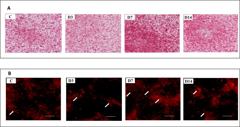
Figure 3. Representative images of collagen deposition at Day 14 of culture.
(A) Representative images of control cells (C), and cells exposed to three (D3), seven (7D), and 14 (D14) days of ES stained with Sirius Red at Day 14 of culture. (B) Stained collagen showed by fluorescence microscopy. Arrows indicate collagen fibers highly condensed in D7 and D14 cells. A lower amount of fibers was observed in D3 and control cells.

