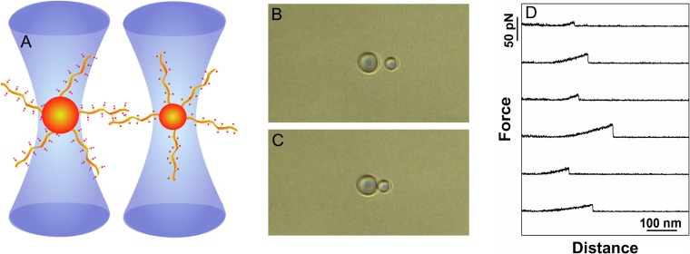
Fig. 2.
Schematic illustration of quantitative determination of carbohydrate–carbohydrate interactions employing dual beam optical tweezer. (A): A polystyrene bead functionalized with glycans or glycosylated molecules (the example depicts mucins of different but well-defined glycosylation pattern) is trapped in each of the two optical traps of the dual beam optical tweezers instrument. During the experiment, the two beads are brought into contact, allowing the molecules immobilized onto the bead surfaces to interact, before being separated. (B) and (C): Optical micrographs of the two optically trapped beads prior to (B) and in contact (C). The net displacements of the polystyrene beads from the center of the calibrated optical trap are continuously recorded and used for quantifying the force acting on the beads. (D): Examples of force-distance curves determined when separating two mucin-functionalized polystyrene beads. The selected force-distance curves reveal force jumps reflecting the rupture of CCIs formed between the immobilized mucin molecules.

