Figure 4.
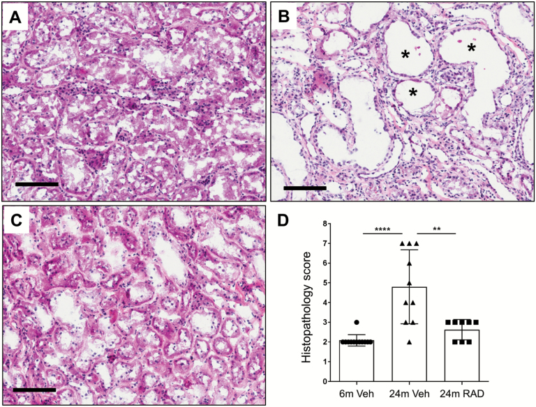
Representative photomicrographs illustrating longitudinal sections of hematoxylin and eosin (H&E)-stained kidneys (cortex region) from a 6-month-old rat treated with vehicle (A), a 24-month-old rat treated with vehicle (B) and a 24-month-old rat treated with RAD001 (C). Asterisks (*) in panel B indicate dilated renal tubules in the kidney from a 24-month-old rat treated with vehicle. Scale bars, 100 μm. (D) Graphical summary of semiquantitative histopathology scores. Data are mean ± SD. ****p ≤ .0001, **p≤ = .01. N = 8–10 rats per group.

