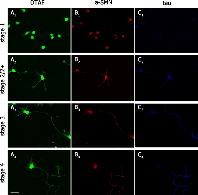
Fig 1. a-SMN subcellular localization during neuronal differentiation.
Confocal images of primary hippocampal neurons from embryonic rats co-labeled with DTAF; (A1-A4, green), rat-specific anti-a-SMN antibody #553 (B1-B4, red) and axonal marker anti-tau antibody (C1-C4, blue). Note the early a-SMN staining within cell bodies in stage 1 (B1) and newly formed primary neurites in stage 2/2+ (B2), and the more selective staining of the forming primary axon in stages 2+-4 (B3-4), with a distribution similar to tau axonal staining (C2-C4). Scale bar: 25 μm.

