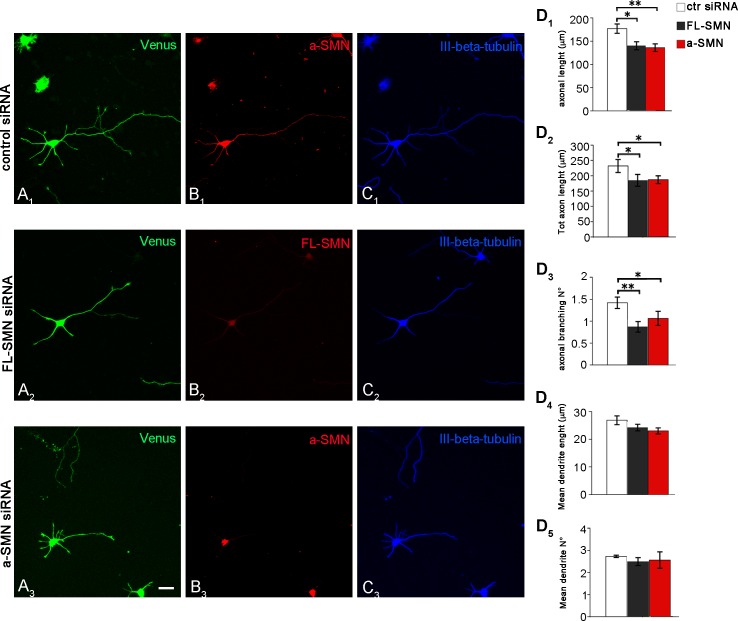
Fig 5. Effect of FL-SMN and a-SMN knockdown on axon growth.
Confocal images of stage 3 hippocampal neurons co-transfected with the Venus plasmid (green) and control (A1-C1) or FL-SMN (A2-C2) or a-SMN specific siRNAs (A3-C3), labeled with anti-a-SMN (red, B1 and B3), anti-SMN (red, B2), and III-ß-tubulin (blue, C1-C3) antibodies. Note that, if compared with neurons treated with control siRNA, both FL-SMN- and a-SMN silenced hippocampal neurons showed after 3 DIV shorter and less extensively branched axons. (D) Morphometric analysis revealed that FL-SMN or a-SMN knock-down were equally effective in reducing axon elongation (FL-SMN and a-SMN vs. control siRNA: * p < 0.05, ** p < 0.01: D1), total axon length (FL-SMN and a-SMN vs. control siRNA: * p < 0.05: D2) and axonal branching (FL-SMN and a-SMN vs. control siRNA: * p < 0.05, ** p < 0.01: D3). By contrast, dendrite length (D4) and number (D5) were unaffected by either FL-SMN or a-SMN knock-down. Data are presented as mean ± SEM of three independent experiments (axon elongation n > 250 cells, total axon length and axonal branching n > 140 cells, dendrite length and number n > 240 cells). Statistical analysis were performed by means of one-way ANOVA followed by Tukey HSD as post hoc comparison test. Scale bar: 10 μm.

