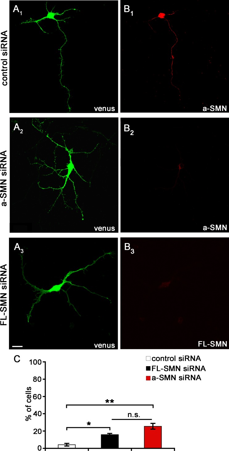
Fig 6. FL-SMN and a-SMN knockdown: Multi-neurite neurons.
(A1-B3) Confocal images of hippocampal neurons co-transfected with control or a-SMN or FL-SMN-specific siRNAs together with Venus plasmid (green, A1-A3), fixed after 3DIV and labeled with the anti-a-SMN #553 (red, B1-B2) or anti-FL-SMN antibodies (red, B3). After selective a-SMN or FL-SMN silencing, a significant fraction of hippocampal neurons showed several processes with similar length and no clear evidence of axonal polarization. (C) Percentages of Venus+ hippocampal neurons displaying multi-neuritc morphology after transfection with control (white bars), FL-SMN SiRNA (grey bars) or a-SMN-specific siRNAs (red bars). Data are presented as mean ± SEM of three different experiments of each group (* p < 0.05; ** p < 0.01; n.s.: not significant). Statistical analysis were performed by means of one-way ANOVA followed by Tukey HSD as post hoc comparison test. Scale bar: 10 μm.

