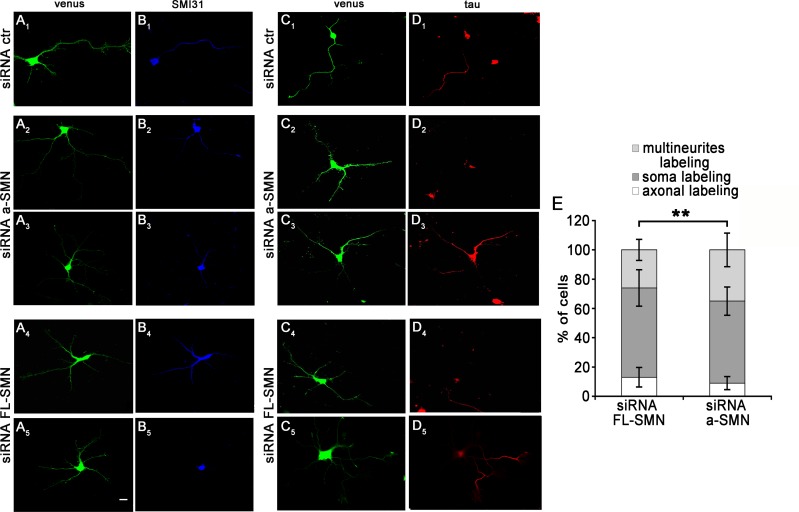
Fig 7. Axon markers SMI31 and tau in multi-neuritic neurons.
(A-D) Confocal images of hippocampal neurons co-transfected with control (A1-D1), a-SMN (A2-D3) or FL-SMN (A4-D5) specific siRNAs and the Venus plasmid (green, A1-A5 and C1-C5), fixed at 3DIV and labeled with the axonal markers SMI31 (blue, B1-B5) or tau (red, D1-D5). While neurons transfected with control siRNA had a single axon labeled by SMI31(A1-B1), in a-SMN and FL-SMN-silenced neurons displaying multi-neuritic morphology, SMI31or tau axonal staining was mainly in soma (B5, D2, D4) or variably localized in two or more neuritic processes (B2-B4, D3, D5). (E) Quantification of axon labeling in individual multi-neuritic neurons. Stacked histograms showing SMI31 distribution in a-SMN and FL-SMN silenced neurons with multi-neuritic morphology. Data are presented as mean ± SEM. At least 70 cells from three different experiments were analyzed. Percent ratio of axonal markers distribution (multineurites/ only soma/axonal labeling) was compared by means of chi-square test (** p<0.01) between the two experimental conditions (a-SMN or FL-SMN siRNA). Scale bar: 10 μm.

