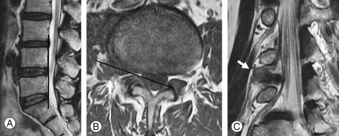
Fig. 1.

Magnetic resonance (MR) images of a 52-year-old female with severe pain in the right leg. Trajectory and angle of oblique lumbar magnetic resonance imaging. (A) Sagittal line of oblique image (black line) is parallel to the line from the posterior margin of the upper to lower end plate of the functional segmental unit in the affected level. (B) Axial angle of oblique image (black line) is parallel to the foramen. Axial T1-weighted MR image indicates ruptured disc particles in the right extraforaminal region of L4–5 (arrow). (C) Lumbar oblique MR image indicate each lumbar nerve root obliquely passing under the pedicles. Right side T2-weighted oblique MR image indicates the cut off of the exiting L4 nerve root by the ruptured disc at the extraforaminal area (arrow).
