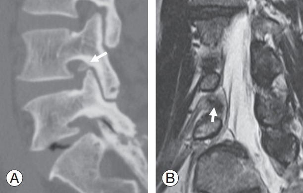
Fig. 4.

Radiologic image of a 56-year-old male with right radiculopathy. (A) Sagittal reconstruction computed tomographic image indicates a bony cyst at the right intervertebral foramen of L4–5 (arrow). (B) Right side T2-weighted oblique magnetic resonance imaging indicates compression of L4 nerve root by bony cyst (arrow).
