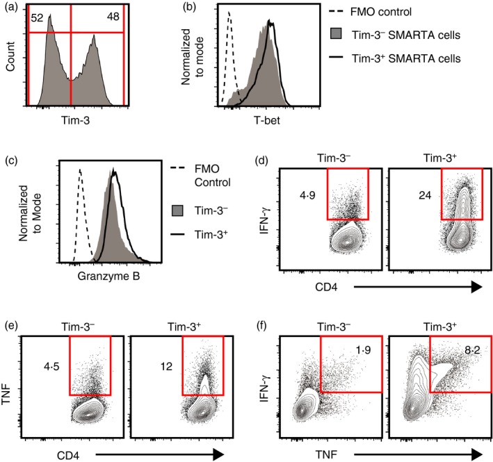
Figure 3.

Differentiation of SMARTA CD4 T cells into Tim‐3− and Tim‐3+ T helper type 1 (Th1) cells. SMARTA CD4 T cells were differentiated under Th1 conditions for 5 days, washed and then cultured under Th1 conditions for another 5 days. Cells were then harvested and analysed. (a) Surface Tim‐3 expression by recovered cells. (b–f) Expression of T‐bet (b), Granzyme B (c), interferon‐γ (IFN‐γ) (d), tumour necrosis factor (TNF) (e), and IFN‐γ and TNF (f) by Tim‐3− and Tim‐3+ cells. For (b) and (c), FMO control represents data from cells stained with all antibodies except those specific for T‐bet or Granzyme B. All data shown are representative of results from at least two independent experiments. [Colour figure can be viewed at http://wileyonlinelibrary.com]
