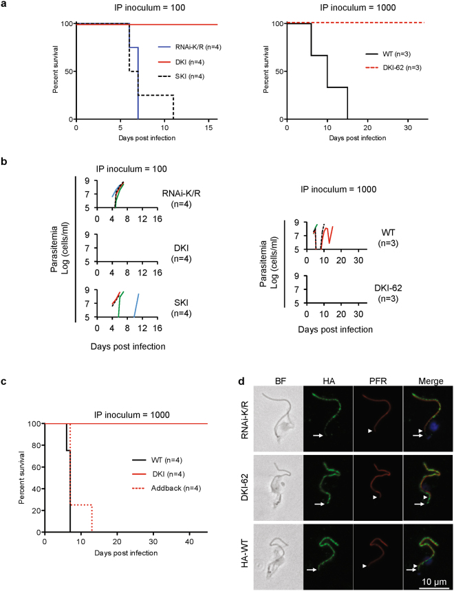
Figure 2.
Virulence defect of LC1 DKI motility mutants. (a) Survival curves for mice infected intraperitoneally with the indicated cell lines. Graphs show data from two independent experiments, RNAi-K/R vs DKI vs SKI (inoculum = 100 parasites, n = 4 mice each) and WT vs DKI-62 (inoculum = 1000 parasites, n = 3 mice each). Data are representative of the phenotypes observed for WT, RNAi-K/R and DKI cell lines across multiple independent experiments (Figs 2c, 3c, and data not shown)17. DKI and DKI-62 are independently derived LC1 K203A/R210A double knock-in mutants. (b) Parasitemia of mice used for panel a. Parasitemia in blood was measured beginning four days post infection. Detection limit is ∼1e5 cells/ml. (c) Survival curves for mice infected intraperitoneally with the indicated cell lines. Addback is a DKI mutant line into which a WT copy of LC1 has been introduced at an ectopic locus, as described in Methods. (d) Immunofluorescence microscopy of detergent-extracted cytoskeletons. HA-tagged LC1 (green), PFR (red), DAPI (blue). The white arrows and arrowheads mark the proximal ends of LC1 and PFR staining, respectively. RNAi-K/R cells were examined 72 h after Tet-induction16,17. Images were collected under identical conditions and processed identically.

