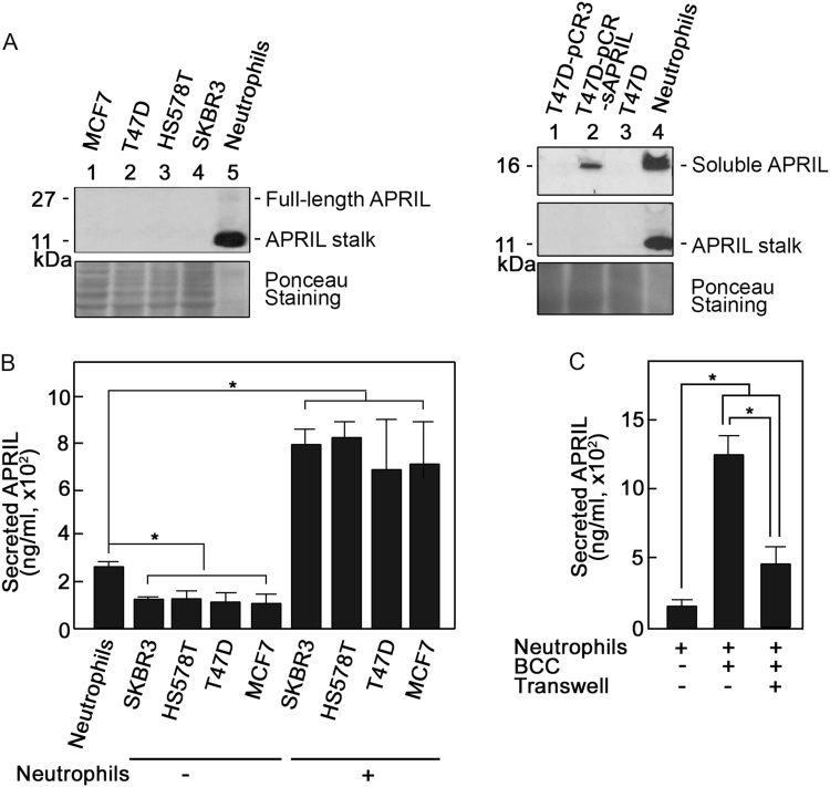
Fig. 1. Breast cancer cells induce secretion of APRIL from neutrophils.
Following approval by the University of Calgary’s Conjoint Health Research Ethics Board, blood was obtained from healthy volunteers that gave informed consent. Neutrophils were isolated as described previously48,49 and suspended in Hank’s Balanced Salt Solution with Ca2+/Mg2+ (Gibco). BCCs (Hs578T, SKBR3, T47D, and MCF7) obtained from the American Type Culture Collection (not authenticated but tested for mycoplasma in the current year) were cultured in Dulbecco’s Modified Eagle Medium (Gibco) with 10% fetal bovine serum (Gibco) and 100 U/ml each of penicillin and streptomycin (Gibco) in 5% CO2 at 37 °C. a APRIL stalk is clearly detectable in neutrophils but not in breast cancer cells. On the left panel, lysates of MCF7, T47D, Hs578T, and SKBR3 cells (60 μg each), as well as neutrophils (5 μg) were subjected to SDS-PAGE and western blotting using APRIL stalk antibody (ED, MyBioSource: MBS194153). On the right panel, neutrophils and T47D cells [non-transfected and transfected with pCR3 (T47D-pCR3) or pCR3-soluble APRIL (T47D-pCR3-sAPRIL)] were immunoblotted using Aprily-5 (Novus Biologicals: NBP1-97599; top panel) and reblotted using April ED (middle panel) antibodies. Aprily-5 detects soluble APRIL. The bottom panel shows Ponceau S staining of the membranes. b Breast cancer cells enhance APRIL secretion from neutrophils. Neutrophils (2 × 10⁶) and breast cancer cells (5 × 10⁵) were incubated alone or together. Cells were then pelleted and supernatants were analyzed for secreted neutrophil APRIL by direct ELISA. Recombinant APRIL (Alexis Corp.) was used as standard. Anti-APRIL (H-60, Santa Cruz Biotech.:sc-28919) used as detection antibody was complexed with HRP-linked anti-rabbit IgG (Cell Signaling). Immunoreactivity was detected using 3,3′,5,5′-tetramethylbenzidine liquid substrate (Sigma). The colorimetric reaction was stopped using 2 M HCl and measured at 490 nm using a SpectraMax M2/M2e multi-mode microplate reader. Measured APRIL in 1 ml corresponds to secretion from 1 × 10⁶ neutrophils. Note that measured levels of APRIL from 5 × 10⁵ and 2 × 10⁶ breast cancer cells were similar and reflect background values. c Breast cancer cell-associated and -released factors are involved in stimulating neutrophil APRIL secretion. Neutrophils (2 × 10⁶) and T47D BCCs (5 × 10⁵) were co-incubated in the presence or absence of transwell membranes (3 μm pore size; Corning) in 24-well plates. When transwell membranes were used, BCCs were loaded in the upper chamber and neutrophils in the lower chamber. Following incubation for 5 min at 37 °C, cells were pelleted and supernatants were analyzed for secreted neutrophil APRIL by ELISA as described above. Values are means ± SD of three replicates of a representative experiment (n = 3). One-tailed Student’s t-test was used to determine statistical significance at p < 0.05. Note that neutrophils used in independent experiments were isolated from different individuals and thus the variation in constitutive/base levels of APRIL secretion and the extent of secretion following stimulation. The specificity of measured APRIL secretion was confirmed using the soluble APRIL blocking peptide, BP150 (data not shown)

