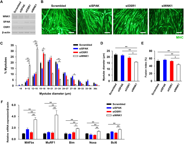
Figure 2.
Small interfering RNA (siRNA)-mediated silencing of WNK1, but not SPAK/OSR1 kinases, induced myotube atrophy and increased atrogene expression. (A) Immunoblots showing the siRNA knockdown efficiency of WNK1, SPAK, or OSR1 in C2C12 cells. (B) After C2C12 cells were pre-treated with siRNA, the cells were incubated in differentiation medium for 4 days, and immunofluorescence study was performed with a myosin heavy chain (MHC) antibody. Scale bar, 200 μm. (C) Histograms showing proportions of myotubes according to the diameters. The proportions of atrophic myotubes were markedly higher in the WNK1-silenced group (n = 5 or 6 per experimental group). (D) The mean myotube diameter value in the WNK1-silenced group was lower than that in the control group (n = 5 or 6 per experimental group). (E) The fusion index represents the percentage of multinucleated MHC-positive myotubes per vision field after 96 h of differentiation (n = 5 or 6 per experimental group). Wnk1-targeted siRNA suppressed the myogenic fusion of myoblasts into myotubes. (F) Quantification of MAFbx, MuRF1, Bim, Noxa, and bcl6 in siRNA-treated C2C12 cells by real-time polymerase chain reaction analysis (n = 4 per experimental group). mRNA levels were normalized against those of β-actin. Values are presented as the mean ± standard error of the mean. *P < 0.05; **P < 0.01. Small interfering RNA, siRNA; WNK, with-no-lysine (K); SPAK, SPS/STE20-related proline-alanine–rich kinase; OSR1, oxidative stress responsive 1; MHC, myosin heavy chain; MAFbx, muscle atrophy F-box; MuRF1, Muscle RING-finger protein-1. Full-length blots are presented in Supplementary Fig. 8.

