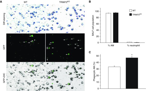
Figure 4.
TRIM72 ablation enhances in vivo AM phagocytosis of P.a. (A) Representative images of BAL fluid (BALF) cell cytospin slides from WT and TRIM72KO mice 1 hour after injection of P.a. GFP. Kwik-diff staining identifies AMs (large, round cells) and neutrophils (star); GFP identifies phagocytic cells (white arrows) and GFP+ differential interference contrast (DIC) identifies internalized GFP bacteria (black arrows) in AMs. (B) Quantification of percentage of AMs and neutrophils in BALF of WT and TRIM72KO mice 1 hour after P.a. injection. (C) Quantification of percentage of phagocytic AM in WT and TRIM72KO mice; n = 3 for each group, *P < 0.05. Data are presented as mean (±SE).

