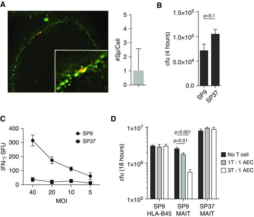
Figure 4.
(A) Primary airway epithelial cells (AECs) were infected with SP9 (multiplicity of infection [MOI] of 5:1) that had been labeled with Alexa Fluor 647 dye and stained with a cell surface mask. At least 100 cells were imaged in three independent experiments, and the number of bacteria per cell was counted using Imaris software (Bitplane). (B) cfu were enumerated by plating serial dilutions from primary AECs infected with SP9 or SP37 for 4 hours. (C) IFN-γ production by D426 G11 in response to SP9- or SP37-infected AECs was measured by ELISPOT assay as previously described. (D) Cells infected with SP9 or SP37 for 4 hours were extensively washed and incubated for an additional 14 hours with the D426 G11 mucosal-associated invariant T (MAIT)-cell clone or the HLA-B45–restricted D466 A10 T-cell clone. cfus were enumerated by plating serial dilutions. Data are representative of four independent experiments.

