Figure 2.
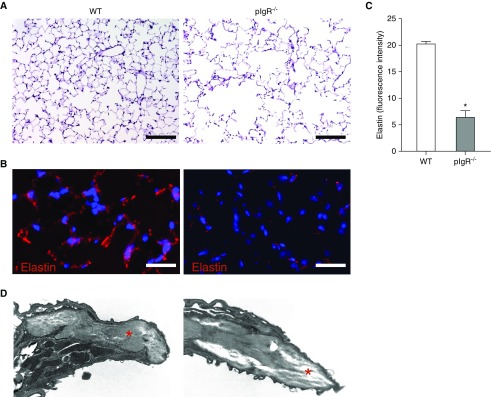
Elastin degradation in lung parenchyma of pIgR−/− mice. (A) Hematoxylin and eosin staining of lung sections shows emphysematous lung destruction in a pIgR−/− mouse (12-mo-old) relative to an age-matched WT control. Scale bars: 50 μm. (B) Immunostaining for elastin from a WT mouse (12-mo-old) and a pIgR−/− mouse. Scale bars: 100 μm. (C) Quantification of fluorescent intensity of elastin staining reported as actual pixel density for each field of lung parenchyma (captured at ×40 objective); n = 6 mice/group; *P < 0.0001 (t test). (D) Transmission electron microscopy image of an interalveolar septum from a WT and pIgR−/− mouse (×50,000). The red stars denote extracellular matrix.

