Figure 6.
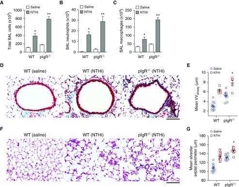
Increased lung inflammation in pIgR−/− mice treated with nontypeable Haemophilus influenzae (NTHi). WT and pIgR−/− mice were treated with NTHi lysates via nebulization once weekly from 2 to 6 months of age. (A–C) Total cells, neutrophils, and macrophages in BAL fluid in WT and pIgR−/− mice treated with NTHi or saline only, as indicated; n = 5–7 mice/group; *P < 0.05 compared with saline-treated WT mice; **P < 0.05 compared with all other groups (ANOVA). (D) Representative images of subepithelial collagen (stained blue) around the small airways of a saline-treated WT mouse or NTHi-treated WT or pIgR−/− mouse as indicated. Masson’s trichrome stain; scale bar: 50 μm. (E) Morphometric analysis of VVairway in WT and pIgR−/− mice as shown in D; n = 5–12 mice/group; *P = 0.05 compared with NTHi-treated WT mice and P < 0.0001 compared with all other groups (ANOVA). (F) Representative images of emphysema in the lungs of a saline-treated WT mouse or NTHi-treated WT or pIgR−/− mouse, as indicated. Hematoxylin and eosin; scale bar: 50 μm. (G) Morphometric analysis of emphysema (mean alveolar septal perimeter) in WT and pIgR−/− mice as shown in F; n = 11–12 mice/group; *P < 0.01 compared with all other groups (ANOVA).

